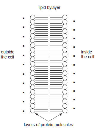
 Heart muscle cells are polarized when at rest. This means that the net
charge density of the fluid inside and outside of the cells is
different, because ion concentrations are different on either side of
the cell membranes. The potential inside of the cells is approximately
-90 mV with respect to the potential outside of the cell membranes. At
rest we find an excess an of positive sodium ions (Na+)
outside of the membranes.
Heart muscle cells are polarized when at rest. This means that the net
charge density of the fluid inside and outside of the cells is
different, because ion concentrations are different on either side of
the cell membranes. The potential inside of the cells is approximately
-90 mV with respect to the potential outside of the cell membranes. At
rest we find an excess an of positive sodium ions (Na+)
outside of the membranes.
A typical cell membrane is relatively impermeable to sodium ions, but
the stimulation of a muscle cell causes an increase in its permeability
to Na+. More sodium ions enter than leave the cell. This
causes a change in the cell potential (depolarization). The potential
inside of the cell becomes positive with respect to the potential
outside of the cell membrane and then returns back to -90 mV, as ions
pumps re-establish the resting potential. The resulting voltage pulse is
called the
action
potential.
In muscle cells, the action potential causes a muscle contraction.
Depolarization and repolarization of the entire heart can be measured on
the skin surface. Such a measurement is called an electrocardiogram
(EKG or ECG). Depolarization of the heart leads to the contraction of
the heart muscles and therefore an EKG is an indirect indicator of heart
muscle contraction.
The cells of the heart will depolarize without an outside stimulus.
This property of cardiac muscle tissue is called automaticity, or
autorhythmicity. The cells responsible for initiating and distributing
the signal to contract are part of the conducting system of the heart, a
network of specialized cardiac muscle cells that initiate and distribute
electrical impulses.
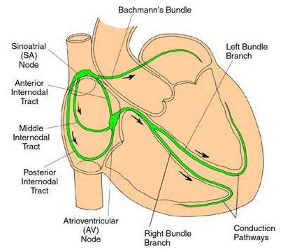 The group of cells that depolarizes fastest is called the
pacemaker or sinoatrial (SA) node.
These cells are located in the
right atrium of the heart. The cells of the atria are all physically connected and
therefore the depolarization of the cells of the SA node causes all the
cells of both atria to depolarize and contract almost simultaneously.
The group of cells that depolarizes fastest is called the
pacemaker or sinoatrial (SA) node.
These cells are located in the
right atrium of the heart. The cells of the atria are all physically connected and
therefore the depolarization of the cells of the SA node causes all the
cells of both atria to depolarize and contract almost simultaneously.
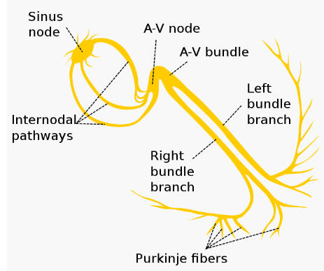
The atria and ventricles are electrically isolated from each other by
connective tissue that acts like the insulation on an electric wire.
Therefore, the depolarization of the atria does not directly affect the
ventricles. However, there is another group of cells in the right atria
called the
AV node that will conduct the depolarization of the atria down a special bundle
of conducting fibers (called the AV bundle or Bundle of His)
to the ventricles.
In the muscle wall of the ventricles are the
Purkinje fibers,
a special system of muscle fibers that bring depolarization to all parts
of the ventricles almost simultaneously. This signal then causes the
ventricular contraction.
There is a time delay inherent in this conduction process starting with
the AV node and atria contraction and ending with ventricular
contraction. This time delay is very important because the atria must
contract before the ventricles. If all the chambers contracted at the
same time, then blood could not flow through the heart because the AV
valves would prevent blood flow between the atria and ventricles.
 When one portion of the heart is polarized and an adjacent portion is
depolarized, then an electrical current moves through the body. This
largest current flows when one half of the heart tissue is polarized and
one half is not polarized. The current decreases when the ratio of
polarized tissue to non-polarized tissue is different from one-to-one.
The change in these current can be measured and displayed. The EKG
represents the sum of all the actions potentials from the heart detected
on the surface of the body. It does not measure the mechanical
contractions of the heart directly.
When one portion of the heart is polarized and an adjacent portion is
depolarized, then an electrical current moves through the body. This
largest current flows when one half of the heart tissue is polarized and
one half is not polarized. The current decreases when the ratio of
polarized tissue to non-polarized tissue is different from one-to-one.
The change in these current can be measured and displayed. The EKG
represents the sum of all the actions potentials from the heart detected
on the surface of the body. It does not measure the mechanical
contractions of the heart directly.
A sample EKG voltage vs. time plot is shown below.
The small P wave accompanies the depolarization of
the atria. The atria begin contracting around 100 ms after the start of the
P wave.
The QRS complex appears as the ventricles depolarize. This
is a relatively strong electrical signal because the mass of the
ventricular muscle is much larger than that of the atria. The
ventricles begin contracting shortly after the R wave.
External links:
AI Study Tip:
Example prompt: How does an EKG measure the electrical activity of the
heart from the surface of the skin? Explain this using the concepts from
our physics module: electric potentials, electric dipoles (specifically how the
heart acts as a shifting dipole), and potential differences measured by
electrodes.